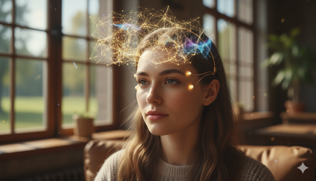
Imagine a toddler gleefully stacking blocks while wearing a colorful helmet, completely unaware she’s offering scientists the clearest picture ever captured of her developing brain. This isn’t a scene from a sci-fi movie—it’s today’s reality, made possible by a seismic shift in neuroimaging powered by wearable technologies. The age of lying motionless inside a claustrophobic MRI tube is rapidly being replaced by an era where mobility, accessibility, and real-world brain mapping redefine neuroscience.
Breaking the Chains: Moving Beyond the Static Scanner
For decades, the cornerstone tools of brain research—functional magnetic resonance imaging (fMRI) and traditional magnetoencephalography (MEG)—offered remarkable insights but came with equally significant limitations. These systems demanded that subjects lie completely still inside massive, immobile machines for extended periods, often in noisy and claustrophobic settings. The cost, complexity, and discomfort of these scans made them particularly unsuitable for children, individuals with neurological conditions, or anyone unable to remain still. Even more critically, the rigid and artificial scanning environment meant that brain activity could only be studied in isolation—divorced from the dynamic, social, and multisensory experiences of everyday life.
Wearable brain imaging technologies have broken through these constraints, offering a revolution in how and where the brain can be studied. By freeing subjects from the confines of traditional scanners, researchers can now observe neural activity as it unfolds in real time during natural behaviors—walking, talking, playing, and engaging socially. This mobility is not just a technical upgrade; it represents a paradigm shift in neuroscience. It allows scientists to capture how the brain truly functions—in motion, in context, and in concert with the world around it—ushering in a new era of discovery that better reflects the lived human experience.
Revolutionary Technology: Quantum Sensors and Light-Based Imaging
At the heart of this transformation are two pioneering technologies: Optically Pumped Magnetometer Magnetoencephalography (OPM-MEG) and High-Density Diffuse Optical Tomography (HD-DOT).
OPM-MEG leverages compact quantum sensors, about the size of LEGO bricks, embedded within lightweight helmets. Developed at the University of Nottingham and commercialized through Cerca Magnetics, these sensors detect the brain’s faint magnetic signals without the need for bulky cryogenic cooling. A groundbreaking matrix coil system actively cancels Earth’s magnetic field, allowing participants to move freely—even walk—while undergoing scans. This innovation marks the first time in history that researchers can observe high-resolution brain activity in motion.
Meanwhile, HD-DOT uses near-infrared light to measure blood flow changes linked to neural activity. Pioneered by research teams such as those at Washington University, the early, cumbersome versions of HD-DOT have been refined into sleek, four-pound caps powered by lightweight backpacks. Companies like EsperImage are leading the way in developing untethered, mobile versions that offer high spatial resolution and reduced sensitivity to movement-related artifacts.
These technologies complement each other. While OPM-MEG excels at detecting the brain’s electrical activity with fine temporal resolution, HD-DOT offers valuable insights into hemodynamic (blood flow) changes with greater spatial precision—making both tools indispensable for wearable brain imaging.
Table 1: Comparing Wearable Brain Imaging Technologies
| Feature | OPM-MEG (e.g., Cerca Magnetics) | HD-DOT (e.g., EsperImage, Optohive) | Traditional fMRI/MEG |
|---|---|---|---|
| Mobility | Full movement (walking allowed) | Limited movement (seated/standing) | No movement |
| Weight/Comfort | Lightweight helmet (~1kg) | Cap (~2-4kg) | Heavy, confined |
| Key Measurement | Magnetic brain fields (Neuronal) | Blood flow (Hemodynamic) | Blood flow (fMRI) / Magnetic fields (MEG) |
| Ideal For | Electrophysiology, Development | Cognitive tasks, Natural interactions | High-res anatomy, Static studies |
| Child-Friendly? | Excellent (Down to infants) | Good | Poor |
Transformative Discoveries: Rewriting the Book on Brain Development
The true power of wearable brain imaging lies not just in its technological elegance but in the transformative discoveries it has enabled—many of which were previously thought to be out of reach. By removing the constraints of traditional scanning environments, researchers can now explore neural processes in real time, across age groups and behaviors, revealing patterns of brain activity that were simply invisible before.
One of the most compelling breakthroughs comes from studies conducted at the University of Nottingham and Toronto’s SickKids Hospital, where scientists have successfully used OPM-MEG to scan children as young as two years old. These pioneering efforts have allowed researchers to map how brain connectivity changes during essential developmental milestones such as language learning, motor coordination, and social engagement. The ability to obtain such detailed and accurate neural data from very young children is establishing new baselines for what typical brain development looks like—baselines that will serve as crucial reference points for identifying atypical patterns early in life.
In a landmark 2025 study, researchers utilized wearable MEG to scan more than 100 individuals ranging from two to thirty-four years old. Their analysis uncovered a critical developmental trajectory: excitatory neural activity tends to decrease with age, while inhibitory signals become stronger, a shift known as the Excitatory-Inhibitory (E-I) Balance. This balance plays a fundamental role in brain maturation and cognitive stability. Disruptions in the E-I Balance have been closely linked to conditions like autism spectrum disorder (ASD), attention deficit hyperactivity disorder (ADHD), and epilepsy. Until now, observing this transition in such a broad and diverse cohort—especially among toddlers—was nearly impossible without the flexibility and safety of wearable neuroimaging.
At SickKids Hospital, Dr. Margot Taylor and her team are using wearable OPM-MEG helmets to study brain function in toddlers diagnosed with ASD as they play and interact naturally. These sessions have opened an unprecedented window into early neurodevelopment, offering real-time insights into how atypical patterns of connectivity and processing emerge in the context of daily life. By shifting from static, clinical observations to dynamic, child-led exploration, researchers are gaining a deeper understanding of the subtle neural signatures that characterize neurodivergent development.
These discoveries are more than academic milestones—they are practical stepping stones toward earlier diagnoses, more targeted interventions, and more effective therapies. As wearable imaging becomes increasingly accessible, it holds the potential to transform clinical care by offering neurologists and developmental psychologists a real-time, non-invasive tool to assess and support each child’s unique neural trajectory. The ability to observe brain development as it unfolds—rather than through fragmented snapshots—promises to reshape how we understand, diagnose, and support neurodiverse populations for generations to come.
New Insights from Light: Mapping Baby Brains in the Real World
One of the most exciting breakthroughs in wearable brain imaging comes from a recent study led by researchers at UCL and Birkbeck, University of London, which revealed how infants as young as five months old process social and emotional stimuli. Using an advanced high-density diffuse optical tomography (HD-DOT) system developed in collaboration with Gowerlabs, a UCL spin-out, the team successfully mapped activity across the entire outer surface of a baby’s brain—a feat previously limited to MRI scanners or small-scale optical systems targeting isolated regions.
Unlike older approaches that focused on only one or two areas at a time, the new headgear allowed for full-head imaging using harmless near-infrared light. Sixteen infants wearing the lightweight cap watched videos mimicking both social interactions—such as actors singing nursery rhymes—and non-social activities like rolling toys. The researchers observed striking patterns: brain activity was more localized during social stimuli, and unexpectedly, the prefrontal cortex lit up—an area involved in emotion and social processing—suggesting that even at five months, babies are already beginning to understand social cues.
Dr. Liam Collins-Jones from UCL emphasized the leap this technology represents: “Instead of viewing the brain in isolated parts, we can now observe widespread activity and interactions across regions in a natural setting.” This capability could lead to earlier detection of neurodevelopmental differences, offering vital insights into conditions such as autism, dyslexia, and ADHD.
Professor Emily Jones, a co-author from Birkbeck, summed up its potential impact: “This wearable technology enables us to observe the baby brain as it plays, learns, and connects with the world—without the restrictive setting of an MRI scanner.”
The study, published in Imaging Neuroscience and presented at the British Science Festival, demonstrates how HD-DOT is becoming a powerful complement to other wearable technologies like OPM-MEG. Both approaches aim to liberate neuroscience from the static scanner, empowering researchers to study the brain in motion, across ages and environments.
Real-World Impact: Clinics, Playing Fields, and Outer Space
The real-world applications of wearable brain imaging are as diverse as they are transformative, extending far beyond research labs into clinics, sports arenas, and even space missions. In clinical neurology, these technologies are opening up new possibilities for understanding and managing movement disorders. For instance, researchers can now monitor brain activity in patients with Parkinson’s disease as they walk, offering unprecedented insights into phenomena like gait freezing. Capturing brain dynamics during actual movement allows clinicians to identify patterns that were previously masked in stationary scans, leading to more personalized and responsive treatment strategies.
In sports medicine, wearable MEG systems are being adapted into lightweight helmets that could one day be worn by athletes during training and competition. These helmets have the potential to detect subtle neural changes that signal incomplete recovery from concussions or early signs of impact-related brain injury. By providing real-time feedback on brain health, such technology could become a crucial tool in protecting athletes from long-term cognitive damage and informing safer return-to-play decisions.
Pediatric care is also undergoing a quiet revolution. Startups like Optohive are developing modular and user-friendly devices such as the OptoCap, designed specifically for clinical use with children. These systems make it possible for clinicians to monitor brain development over time, track rehabilitation progress in kids recovering from brain injury, or customize therapeutic interventions based on a child’s unique neurophysiological profile. Such tools not only make brain imaging more accessible but also bring neuroscience into routine healthcare settings, where early detection and tailored care can have lifelong benefits.
Beyond Earth’s atmosphere, wearable brain imaging is being embraced by space agencies like NASA, which are using near-infrared spectroscopy (NIRS) systems to study astronauts’ brain function in extreme environments. In microgravity or at high altitudes, astronauts can experience cognitive decline, shifts in brain fluid distribution, and psychological stress. Wearable imaging devices allow researchers to track these changes in real time, helping to safeguard brain health during long-duration space missions. This research could also inform our understanding of human cognition under stress here on Earth, from high-altitude pilots to deep-sea explorers.
As these systems become more portable, precise, and intelligent, their ability to monitor brain function across environments and populations positions them not just as research tools—but as powerful instruments of public health, performance optimization, and human exploration.
Challenges Ahead: Shrinking the Tech and Scaling the Science
While wearable brain imaging has made remarkable strides, several critical challenges still stand in the way of its widespread adoption and integration into clinical and everyday settings. Chief among these are technical hurdles related to signal fidelity. Movement artifacts, ambient noise, and electromagnetic interference can all compromise the delicate measurements these devices are designed to capture—especially in uncontrolled environments. Overcoming these issues will require continuous innovation in signal processing, including real-time filtering, artifact rejection, and machine learning algorithms that can distinguish meaningful brain signals from background noise.
Another major challenge lies in the sheer volume and complexity of the data generated. Wearable systems often collect multiple streams of information simultaneously—neural activity, motion, physiological signals like heart rate or respiration—resulting in large, high-dimensional datasets. Extracting actionable insights from this data in a way that clinicians and researchers can use effectively demands the development of robust, intuitive analytical frameworks. AI and data visualization tools will play an essential role in bridging the gap between raw data and real-world decision-making, whether in a hospital, a research lab, or a sports facility.
The path to commercialization presents its own set of obstacles. Devices like EsperImage’s HD-DOT caps, while powerful in research environments, must undergo miniaturization and refinement before they can be manufactured at scale and offered at a cost accessible to hospitals, schools, and rehabilitation centers. Ensuring that these devices retain their precision and reliability during this transition from lab prototype to consumer-grade technology is no small feat, requiring cross-disciplinary collaboration between neuroscientists, engineers, and manufacturers.
Equally important is the need for regulatory validation. Gaining approval for clinical and diagnostic use requires extensive peer-reviewed testing, long-term outcome studies, and compliance with health technology standards. Without formal validation, these technologies will remain confined to research, unable to fulfill their potential to revolutionize healthcare. As the field advances, navigating the regulatory landscape—while preserving innovation—will be essential to moving wearable brain imaging from proof-of-concept to clinical practice.
Conclusion: A New Window Into the Human Experience
Wearable brain imaging represents far more than a technological innovation—it marks a paradigm shift in how we explore the mind. For the first time, we can observe the human brain in context, capturing its activity as a child learns to speak, as friends laugh in conversation, or as an athlete pushes their physical limits. This new window into the brain’s functioning opens unprecedented opportunities for medicine, education, space exploration, and beyond.
“Just five years ago, acquiring high-resolution brain images while someone walked seemed like science fiction,” says Professor Matt Brookes from the University of Nottingham. “Now it’s reality—and we’re just scratching the surface.”
As these devices grow smaller, smarter, and more integrated with AI, they promise not only to revolutionize neuroscience research but also to enhance everyday healthcare. The static snapshots of yesterday are giving way to dynamic, real-time recordings of the brain in motion. We are entering a new era of neuroscience—one where the human experience can finally be understood as it truly unfolds: in motion, in interaction, and in life.
International Defense Security & Technology Your trusted Source for News, Research and Analysis
